Technology
The Department of Radiation Medicine provides access to state-of-the-art treatment, planning, and imaging systems across our clinics. The systems below are a sample of what we currently use to care for patients and train the next generation of radiation oncologists and medical physicists.
Newest at UW
Recently added systems that expand our treatment, planning, and imaging capabilities.
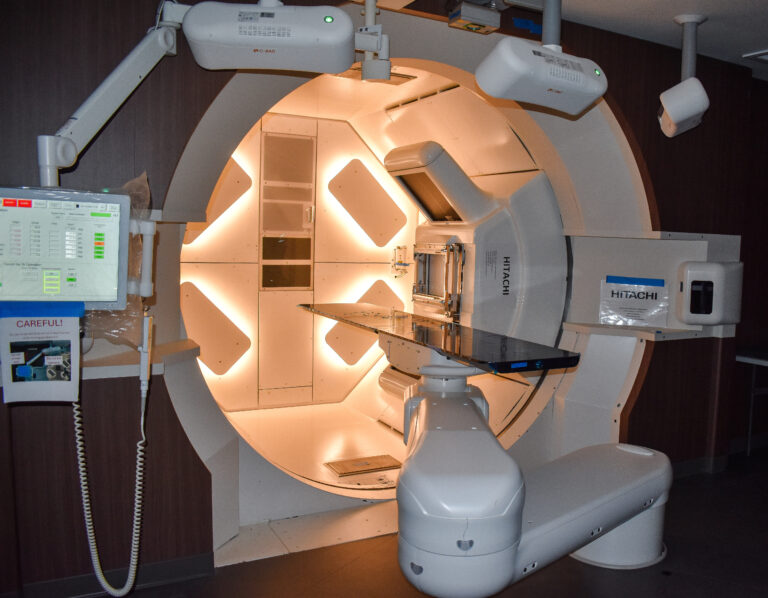
A proton beam treatment that targets tumors precisely while sparing nearby healthy tissue. Both gantry-based and fixed-beam configurations deliver highly conformal dose distributions with minimal exit dose, well suited to complex tumors near critical structures.
Location: EastPark Medical Center
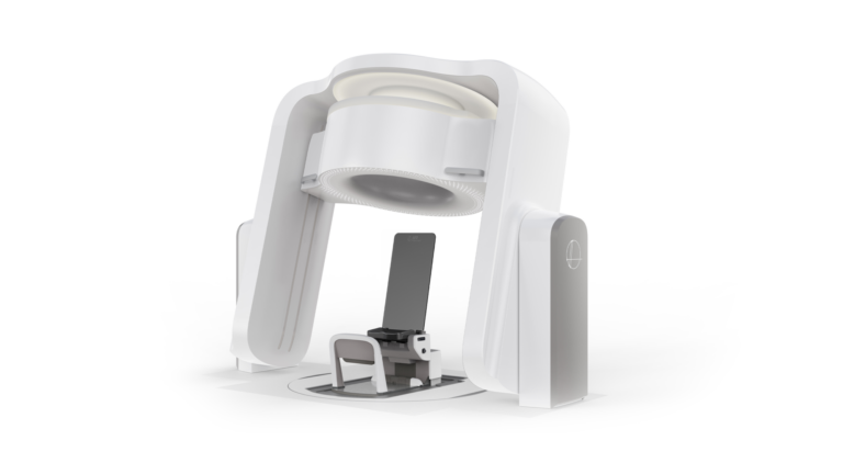
Leo Cancer Care Marie® upright CT photon therapy
An upright CT scanner that images patients sitting upright instead of lying down. Upright positioning improves comfort and anatomical alignment, particularly for thoracic and head-and-neck cases, and supports future upright proton therapy workflows.
Location: EastPark Medical Center
RayCare treatment planning system
An oncology information system that connects planning, scheduling, and patient data in one place. Integrates with RayStation to provide adaptive planning, workflow automation, and multi-modality treatment support including proton therapy.
Location: EastPark Medical Center
Treatment delivery systems
The machines that deliver radiation to patients. Each system is suited to different cancer types and treatment approaches.
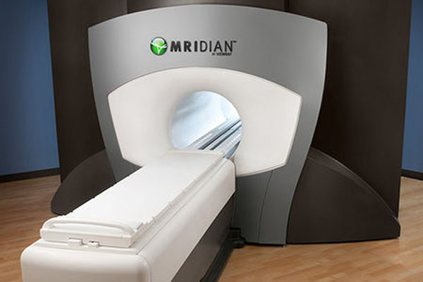
Combines a linear accelerator with real-time MRI imaging so clinicians can see tumors during treatment. Real-time MRI imaging and adaptive planning during therapy sessions, allowing dose adjustments based on the day’s anatomy.
Location: University Hospital
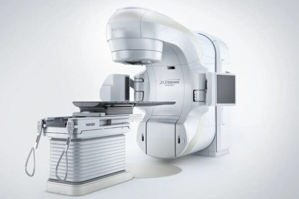
Varian TrueBeam
A versatile linear accelerator used across many cancer types and treatment techniques. Features VisionRT AlignRT optical surface tracking, PerfectPitch 6 degrees of freedom couches, and multiple photon and electron beam energies.
Location: University Hospital, EastPark Medical Center, Johnson Creek
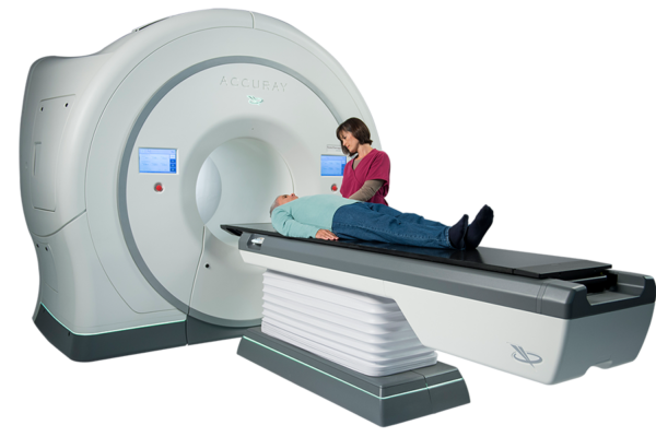
Accuray TomoTherapy HDA
An image-guided radiation therapy platform originally developed here at UW–Madison. Designed for precision and adaptability, the TomoTherapy HDA was developed by physicist Thomas “Rock” Mackie and his team at UW–Madison, and remains a key part of our treatment portfolio.
Location: University Hospital
Elekta Flexitron remote afterloader
A brachytherapy system that places a radioactive source directly inside or next to the tumor for a short time. Delivers a precise, localized dose of radiation while minimizing exposure to surrounding healthy tissue.
Treatment planning systems
The software our physicians and physicists use to design each patient’s treatment plan before delivery.
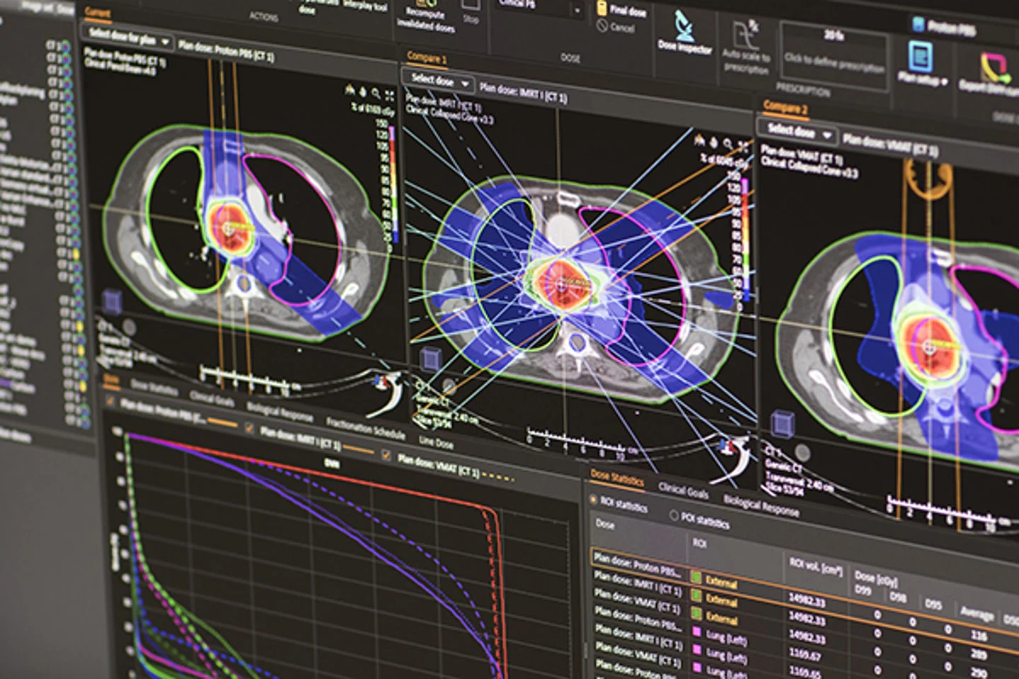
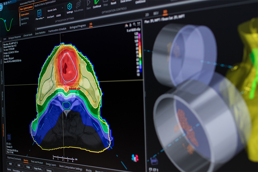
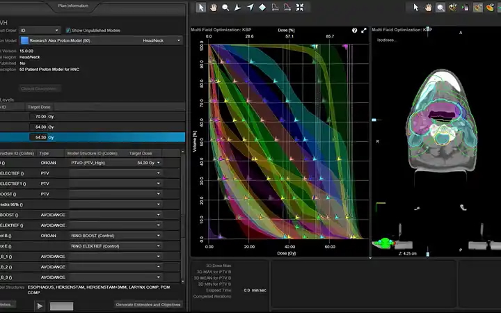
RayStation is a comprehensive treatment planning system by RaySearch Laboratories. It supports external beam therapy including adaptive therapy capabilities, multi-criteria optimization, and advanced automation and scripting.
Eclipse
Eclipse is a treatment planning system by Varian. Here we use Eclipse as our primary platform for stereotactic radiosurgery (SRS) treatment planning, enabling highly precise and conformal dose delivery for intracranial targets. Eclipse supports advanced techniques such as conformal arcs, volumetric modulated arc therapy (VMAT), and cone planning which are critical for achieving steep dose gradients and optimal normal tissue sparing.
ViewRay
The ViewRay treatment planning system for the MRIdian MR-Linac integrates real-time MRI guidance with adaptive radiation therapy, enabling clinicians to visualize soft tissue anatomy during treatment. It supports on-table adaptive planning, allowing dose adjustments based on daily anatomy changes, which improves precision and reduces exposure to healthy tissue.
Oncentra
Oncentra is a specialized treatment planning system for brachytherapy from Elekta. It provides advanced tools for contouring, implant reconstruction, and applicator modeling across for a variety of clinical applications, including gynecologic, breast, and prostate treatments.
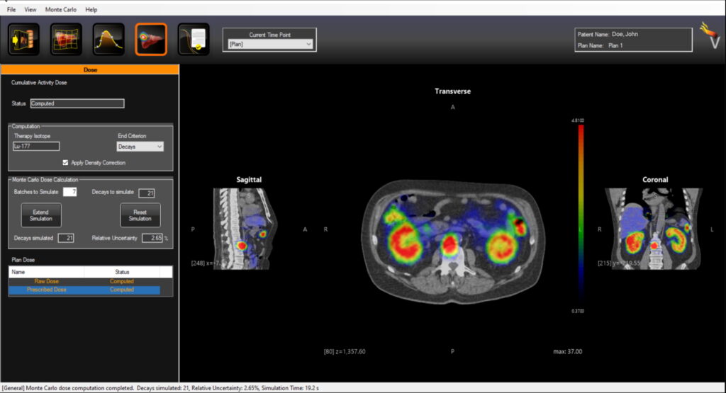
Torch (Voximetry)
The Torch (Voximetry) is GPU-based Monte Carlo dosimetry software designed for personalized radiopharmaceutical therapy (RPT). It supports workflows from DICOM image import (SPECT/CT, PET/CT) through image registration, ROI segmentation, pharmacokinetic modeling, and absorbed-dose calculation per voxel for a variety of radiopharmaceuticals. At UW, Torch is used to calculate patient-specific dosimetry for treatments like Lu-177 vipivotide tetraxetan (Pluvicto®) and Lu-177 dotatate (Lutathera®).
Advanced imaging and guidance systems
CT, MRI, and ultrasound systems that locate tumors precisely before and during treatment. Grouped by what they do, with models listed by clinic.
Siemens CT Simulators
Where patients are scanned to plan their radiation treatment. All three simulators offer advanced reconstruction, multi-energy scanning, and motion management including breath hold and 4DCT scanning with the Varian RGSC respiratory gating system.
Learn more about Siemens Healthineers technology
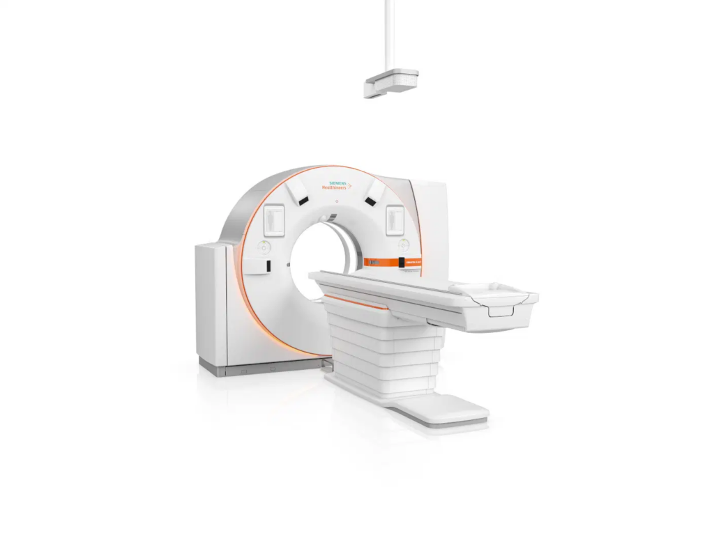
Siemens SOMATOM X.ceed
Location: EastPark Medical Center
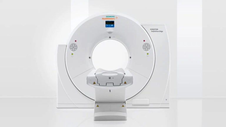
SiemensEdge
Location: University Hospital
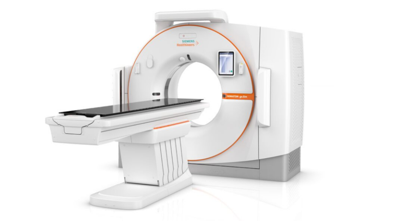
Siemens.Go
Location: Johnson Creek
CT for brachytherapy
CT imaging that happens during the procedure, so the patient doesn’t have to be moved.
Both systems enable real-time verification of applicator placement, target geometry, and organ-at-risk positioning, supporting adaptive brachytherapy planning and intra-procedural decisions.
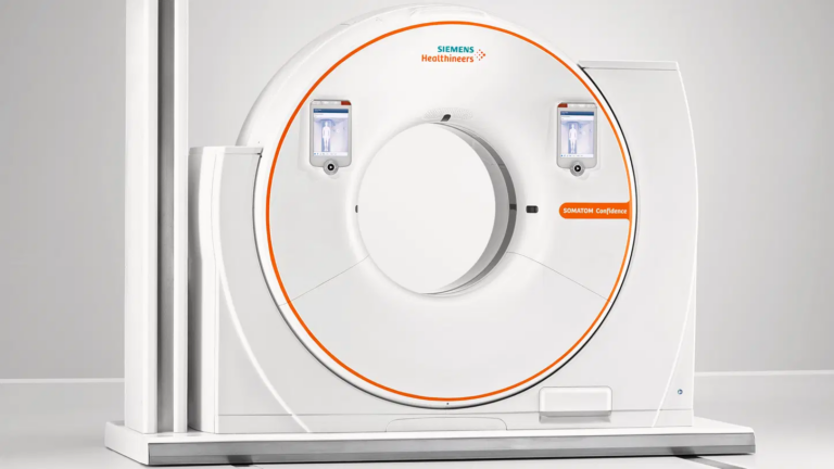
Siemens Confidence CT-on-Rails
The Siemens Confidence CT-on-Rails located in our EastPark brachytherapy suite allows high-quality CT imaging to be performed with the patient in the treatment position, without the need for patient transport, improving both accuracy and efficiency. The system enables real-time verification of applicator placement, target geometry, and organ-at-risk positioning, supporting adaptive brachytherapy planning and intra-procedural decision-making.
Location: EastPark Medical Center
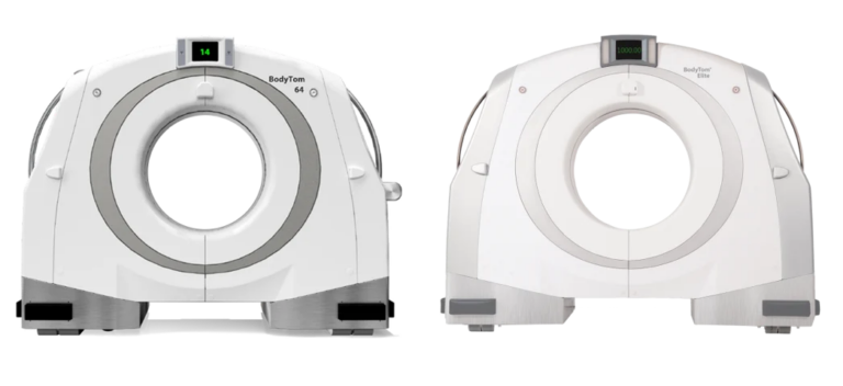
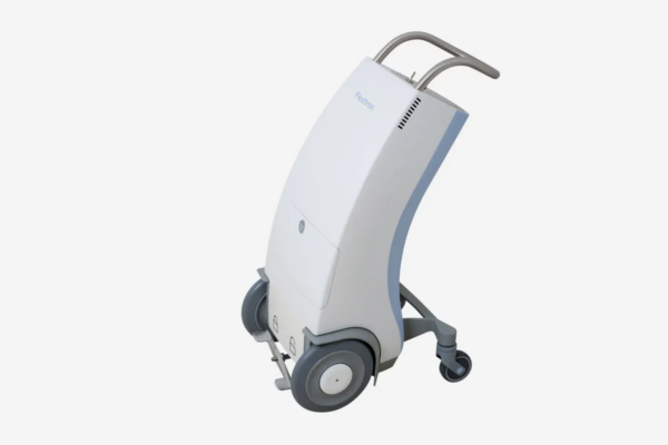
Neurologica BodyTom CT-on-Wheels
The Neurologica BodyTom CT-on-wheels located in our UH brachytherapy suite allows high-quality CT imaging to be performed with the patient in the treatment position, without the need for patient transport, improving both accuracy and efficiency. The system enables real-time verification of applicator placement, target geometry, and organ-at-risk positioning, supporting adaptive brachytherapy planning and intra-procedural decision-making.
Location: University Hospital
MR simulators
High-contrast MRI scanners used to plan treatments where soft tissue detail matters. Having MR simulators directly in the department streamlines scheduling, reduces patient transport, and allows simulation in treatment immobilization for accurate geometric consistency between imaging and delivery.
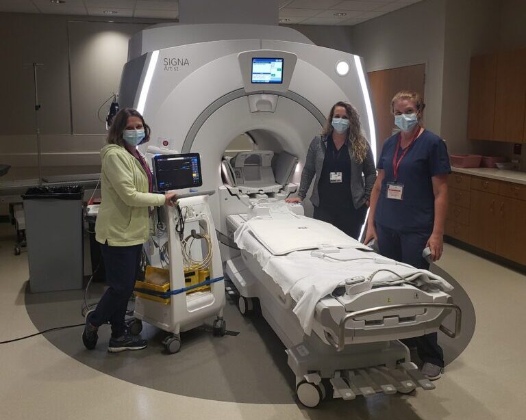
GE Healthcare 1.5 T MRI (EMC/UH)
GE’s 1.5T MRI systems provide provides high-quality soft-tissue–contrast imaging to support advanced radiation therapy planning. Having MR-simulators directly within the department streamlines the clinical workflow, reducing scheduling complexity, minimizing patient transport, and enabling tighter coordination between simulation, contouring, and planning teams. Importantly, patients can be also easily be simulated in their treatment immobilization, ensuring accurate geometric consistency between imaging and delivery.
Location: EastPark Medical Center, University Hospital
Ultrasound
Real-time imaging used to guide brachytherapy needles and applicators. High-resolution imaging supports accurate placement, target localization, and assessment of surrounding anatomy. Primarily used for skin and prostate brachytherapy.
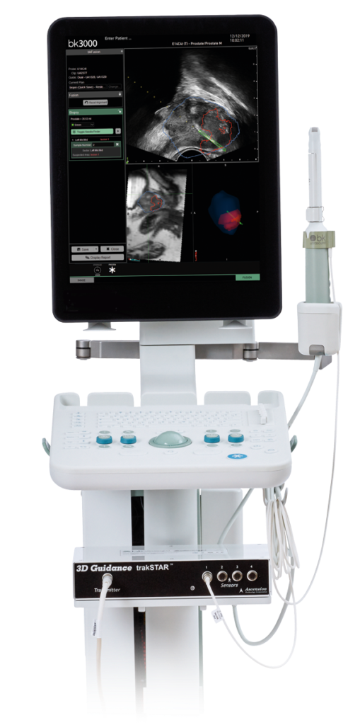
BK Medical Ultrasound
BK Ultrasound systems provide high-resolution, real-time ultrasound imaging to support image-guided brachytherapy procedures. Ultrasound is primarily used for treatment guidance during skin and prostate brachytherapy, where real-time visualization is critical for accurate applicator or needle placement, target localization, and assessment of surrounding anatomy.
Location: All sites
Other software we use
The software our physicians and physicists use to design each patient’s treatment plan before delivery.
SunCHECK by Sun Nuclear is a comprehensive quality management platform for radiation therapy, built on a centralized dashboard and cloud-enabled architecture. We use SunCHECK to perform, document, and track daily, monthly, annual, and post-service quality assurance (QA) across nearly all treatment machines at all of our clinics. This unified system supports standardized QA workflows, improves consistency and traceability, and enables efficient oversight of machine performance and compliance across our multi-site network.

Mobius 3D
Mobius3D is an independent 3D dose verification system designed for radiation therapy quality assurance.It recalculates patient treatment plans using the same DICOM data from the TPS and compares results against the original plan, providing fast, automated checks for dose accuracy and compliance with clinical standards.

MIM Software
MIM Software is an advanced imaging platform used for ROI segmentation, multi-modality image registration, and adaptive dose assessment.